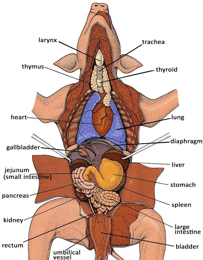
44 digestive system diagram with labels and functions
Organs in the Excretory System and Their Functions Kidneys are bean-shaped organs of a reddish brown color that are found in the sides of the vertebral column. Once the body has extracted what it needs from food and drink, it sends the wastes to the kidneys. The kidneys filter the wastes, including urea, salt and excess water, which are flushed out of the body as urine. 2. Skin The MSDS: A Basic Guide for Users - Canadian WHMIS Version Introduction. A Material Safety Data Sheet (MSDS) provides basic information on a material or chemical product. It contains information on the properties and potential hazards of the material, how to use it safely, and what to do if there is an emergency. This document is intended primarily as a guide for non-technical people who use MSDSs.
Parts and Components of Human Ear and Their Functions The fluid in the ear also helps the body maintain a sense of balance so the body can maintain proper posture and coordination. There are three major parts of the ear, the outer, middle and inner ear. Each contains several parts that are essential to the overall function of the ear.

Digestive system diagram with labels and functions
Lysosome - National Human Genome Research Institute Home Definition A lysosome is a membrane-bound cell organelle that contains digestive enzymes. Lysosomes are involved with various cell processes. They break down excess or worn-out cell parts. They may be used to destroy invading viruses and bacteria. 16 Female Reproductive System Packet Answers energy of a reaction is. Label the following picture: 5. Add the labels to the diagram of the reproductive system of a … Insect Life Cycle: Introduction, Life Cycle, FAQs - BYJUS 08/11/2019 · Number the answers correctly according to the numbering system used in this question paper. Present your answers according to the instructions of each ... Cell Membrane (Plasma Membrane) - Genome.gov The cell membrane, also called the plasma membrane, is found in all cells and separates the interior of the cell from the outside environment. The cell membrane consists of a lipid bilayer that is semipermeable. The cell membrane regulates the transport of materials entering and exiting the cell. If playback doesn't begin shortly, try ...
Digestive system diagram with labels and functions. Biology Archive | May 04, 2022 | Chegg.com Biology Archive: Questions from May 04, 2022. Explain the domains of life, including the number of domains, names of the domain, basic types of organisms found in each domain, and the general type of cells that make up those organisms. DEFINE. Common Diseases of the Circulatory System | MD-Health.com Arteriosclerosis is a common disease of the circulatory system caused by the buildup of fat, cholesterol, or other substance in the artery wall. Deposits in the artery cause the vessel to stiffen and narrow. Diabetes, high cholesterol, smoking, and high blood pressure can result in stiff arteries that restrict blood flow through the heart. 8 Essential Skin Functions - New Health Advisor The cells within the skin like Langerhans cells, phagocytic cells, and epidermal dendritic cells help with immunity. 5. Permit Movement and Growth The skin allows for bodily growth and adapts to suit an individuals course of movement. It has elastic and recoil properties on all of its layers, meaning it can adapt for growth and movement. 6. › resource › t2-s-788-new-humanOrgan Map | Diagram of Human Body Internal Organs Functions Featuring an accurate illustration and informative, clear labels, this organ map is a fantastic visual aid to support your teaching during science lessons all about the human body's internal organs.Once downloaded, you'll have an A4 diagram of a human body and internal organs, clearly labelled and perfect for individual use. Each internal organ is labelled and includes a definition of each ...
Human brain - Wikipedia The main functions of the frontal lobe are to control attention, abstract thinking, behaviour, problem solving tasks, and physical reactions and personality. The occipital lobe is the smallest lobe; its main functions are visual reception, visual-spatial processing, movement, and colour recognition. en.wikipedia.org › wiki › DopamineDopamine - Wikipedia The bulk of this dopamine sulfate is produced in the mesentery that surrounds parts of the digestive system. The production of dopamine sulfate is thought to be a mechanism for detoxifying dopamine that is ingested as food or produced by the digestive process—levels in the plasma typically rise more than fifty-fold after a meal. Gastrointestinal Diagram - non contrast enhanced ct abdominal ct shows ... Gastrointestinal Diagram. Here are a number of highest rated Gastrointestinal Diagram pictures on internet. We identified it from obedient source. Its submitted by admin in the best field. We say you will this nice of Gastrointestinal Diagram graphic could possibly be the most trending topic when we allowance it in google pro or facebook. An Introduction to the 5 Layers of Abdominal Wall - New Health Advisor There are superficial nerves and vessels that go between these two layers. 5. Skin Skin is the outermost layer of the abdominal wall. It protects us from microbes and the elements, helps regulate body temperature, and permits the sensations of touch, heat, and cold. Nerves That Run to the Muscles and Skin of Abdomen
Mechanical and Chemical Digestion - New Health Advisor In the mouth, both mechanical and chemical digestion takes place. 2. Pharynx The pharynx is the place where food is swallowed. The pharynx is the part of the digestive tract that leads to the esophagus. The epiglottis is located within the pharynx. Its job is to close so that food doesn't enter the trachea during the act of swallowing. 3. Esophagus What Is Negative Feedback Loop of Blood Pressure? - New Health Advisor First is a sensor, next is the integrating center, and then the effector (Figure 1). They work together like this: Sensor They sense changes in your body and send a message to the integrating center. Integrating Center It receives messages from the sensors and decides which effector needs a signal to go out and fix the issue in the body. Effectors › type › science-diagramsScience A-Z Science Diagrams - Visual Teaching Tools Science Diagrams from Science A-Z provide colorful, full-page models of important, sometimes complex science concepts. Science Diagrams, available in both printable and projectable formats, serve as instructional tools that help students read and interpret visual devices, an important skill in STEM fields. Brain Structures and Their Functions | MD-Health.com The limbic system includes the amygdala, hippocampus, hypothalamus and thalamus. Amygdala: The amygdala helps the body responds to emotions, memories and fear. It is a large portion of the telencephalon, located within the temporal lobe which can be seen from the surface of the brain. This visible bulge is known as the uncus.
ecampusontario.pressbooks.pub › lymphatic-systemLymphatic and Immune Systems – Building a Medical Terminology ... The lymphatic system is a series of vessels, ducts, and trunks that remove interstitial fluid from the tissues and return it the blood. The lymphatic vessels are also used to transport dietary lipids and cells of the immune system. Cells of the immune system, lymphocytes, all come from the hematopoietic system of the bone marrow.
Respiratory System Organs and Their Functions - New Health Advisor The airways (nose, mouth, pharynx, larynx etc.) allow air to enter the body and into the lungs. The lungs work to pass oxygen into the body, whilst removing carbon dioxide from the body. The muscles of respiration, such as the diaphragm, work in unison to pump air into and out of the lungs whilst breathing. Physiology of Gas Exchange
Emissary veins: Definition, location, function | Kenhub The function of the emissary veins is to provide selective cooling of the brain, as well as an alternative drainage route of the brain in the case of obstruction of dural venous sinuses. Furthermore, their clinical significance is reflected by the fact that they provide a pathway for the spreading of extracranial infections to the neurocranium.
Organs in 9 Abdomen Regions | New Health Guide Umbilicus, Jejunum, Ileum, Duodenum. Right Iliac Fossa. Appendix, Cecum. Left Iliac Fossa. Descending Colon, Sigmoid Colon. Hypogastrium. Urinary Bladder, Sigmoid Colon, Female Reproductive Organs. Below is a reference video, which explains the differential diagnosis of abdominal pain according to the abdominal region.
Sponge - Wikipedia 5 Vital functions 5.1 Movement 5.2 Respiration, feeding and excretion 5.3 Carnivorous sponges 5.4 Endosymbionts 5.5 "Immune" system 5.6 Reproduction 5.7 Coordination of activities 6 Ecology 6.1 Habitats 6.2 As primary producers 6.3 Defenses 6.4 Predation 6.5 Bioerosion 6.6 Diseases 6.7 Collaboration with other organisms 6.8 Sponge loop
Earthworm Dissection - Biology Course Information The dorsal blood vessel and digestive tract should be exposed at this point. Internal View, Crop and Gizzard Labeled Crop and Gizzard Examine the digestive tract . From the mouth back, locate the pharynx, esophagus ( eso = within, inward; phago = to eat), crop , gizzard, and intestine. Refer to these photos, and the large earthworm model.
Cross Section Jellyfish En Clip Art at Clker.com - vector clip art online, royalty free & public ...
quizlet.com › 553341254 › week-15-digestive-systemWeek 15: Digestive System Physiology Flashcards - Quizlet The normal osmolarity of intracellular and extracellular fluid is 300 mOsm, which makes them isotonic. Normal cell and plasma osmolarity is best illustrated by diagram number 1. Mrs. F's liver is no longer producing enough plasma protein so her blood has become hypotonic to her cells. This is best illustrated by diagram number 3.
› consumers › consumer-updatesConsumer Updates | FDA Science-based health and safety information you can trust.
› digestive-systemDigestive System | BioNinja There are two major groups of organs which comprise the human digestive system: The alimentary canal consists of organs through which food actually passes (oesophagus, stomach, small & large intestine) The accessory organs aid in digestion but do not actually transfer food (salivary glands, pancreas, liver, gall bladder) Diagram of the ...

Human Digestive System Labeled - Health Picture Reference | Digestive tract diagram, Human ...
Cell Membrane (Plasma Membrane) - Genome.gov The cell membrane, also called the plasma membrane, is found in all cells and separates the interior of the cell from the outside environment. The cell membrane consists of a lipid bilayer that is semipermeable. The cell membrane regulates the transport of materials entering and exiting the cell. If playback doesn't begin shortly, try ...
16 Female Reproductive System Packet Answers energy of a reaction is. Label the following picture: 5. Add the labels to the diagram of the reproductive system of a … Insect Life Cycle: Introduction, Life Cycle, FAQs - BYJUS 08/11/2019 · Number the answers correctly according to the numbering system used in this question paper. Present your answers according to the instructions of each ...
Lysosome - National Human Genome Research Institute Home Definition A lysosome is a membrane-bound cell organelle that contains digestive enzymes. Lysosomes are involved with various cell processes. They break down excess or worn-out cell parts. They may be used to destroy invading viruses and bacteria.


Post a Comment for "44 digestive system diagram with labels and functions"