40 cell structure with labels
A Labeled Diagram of the Plant Cell and Functions of its Organelles A Labeled Plant Cell. Amyloplasts. A major component of plants that are starchy in nature, the amyloplasts are organelles that store starch. They are classified as plastids, and are also known as starch grains. ... It is a membrane-bound structure that contains the cells hereditary information. Function: Controls expression and transcription of ... Label the Animal Cell: Level 1 | Worksheet | Education.com Label the Animal Cell: Level 1. Students demonstrate their knowledge of the structure of animal cells in this middle grades life science worksheet! In Label the Animal Cell: Level 1, students will use a word bank to label the parts of a cell in an animal cell diagram. To take the learning one step further, have students assign a color to each ...
Label the cell structure. | Study.com Answer to: Label the cell structure. Cells: All living cells contain an intracellular space called the cytoplasm.The cytoplasm is filled with a jelly-like fluid where many of the cells enzymatic ...
Cell structure with labels
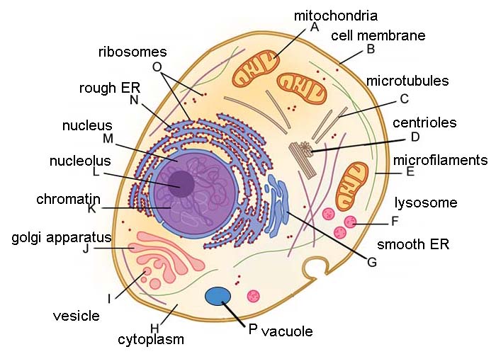
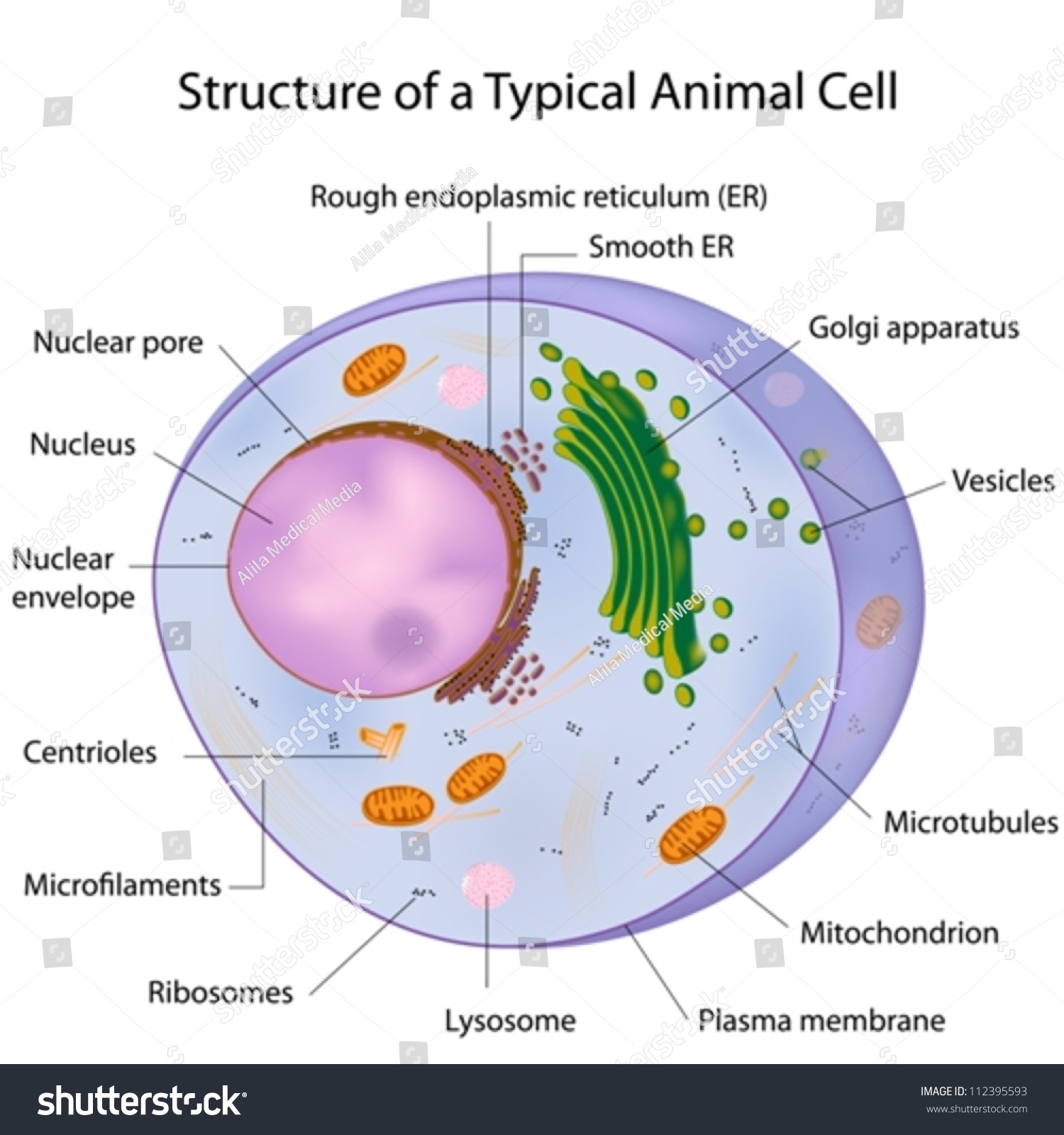
Animal Cell - Structure, Function, Diagram and Types The cell is the structural and functional unit of life. These cells differ in their shapes, sizes and their structure as they have to fulfil specific functions. Plant cells and animal cells share some common features as both are eukaryotic cells. However, they differ as animals need to adapt to a more active and non-sedentary lifestyle. Plant Cell Structure and Parts Explained With a Labeled Diagram Different Parts of a Plant Cell. Plant cells are classified into three types, based on the structure and function, viz. parenchyma, collenchyma and sclerenchyma. The parenchyma cells are living, thin-walled and undergo repeated cell division for growth of the plant. They are mostly present in the leaf epidermis, stem pith, root and fruit pulp. Structure of Cell: Definition, Types, Diagram, Functions - Embibe Structure of Cell: Cell is the basic functional unit that makes up all living organisms. All organisms, including ourselves, start life as a single cell called the egg. Cells are small microscopic units that perform all essential functions of life and are capable of independent existence. With the invention of microscopes, many unknown facts ...
Cell structure with labels. en.wikipedia.org › wiki › Nuclear_envelopeNuclear envelope - Wikipedia Nesprin-mediated connections to the cytoskeleton contribute to nuclear positioning and to the cell’s mechanosensory function. KASH domain proteins of Nesprin-1 and -2 are part of a LINC complex (linker of nucleoskeleton and cytoskeleton) and can bind directly to cystoskeletal components, such as actin filaments , or can bind to proteins in ... Cell Membrane 3 D Structure Labeled : Functions and Diagram Friday, May 14th 2021. | Diagram. Cell Membrane 3 D Structure. It protects the integrity of the cell along with supporting the cell and helping to maintain the cell's shape. The cell membrane, also called the plasma membrane, is a thin layer that surrounds the cytoplasm of all prokaryotic and eukaryotic cells, including plant and animal cells. Human Cell Diagram, Parts, Pictures, Structure and Functions Human Cell Diagram, Parts, Pictures, Structure and Functions. The cell is the basic functional in a human meaning that it is a self-contained and fully operational living entity. Humans are multicellular organisms with various different types of cells that work together to sustain life. Other non-cellular components in the body include water ... › books › NBK26857Cell Junctions - Molecular Biology of the Cell - NCBI Bookshelf Specialized cell junctions occur at points of cell-cell and cell-matrix contact in all tissues, and they are particularly plentiful in epithelia. Cell junctions are best visualized using either conventional or freeze-fracture electron microscopy (discussed in Chapter 9), which reveals that the interacting plasma membranes (and often the underlying cytoplasm and the intervening intercellular ...
Cell Structures and Processes - The Biology Corner Learn the parts of animal and plant cells by labeling the diagrams. Pictures cells that have structures unlabled, students must write the labels in, this is intended for more advanced biology students. Bacteria shapes, structure and diagram - Jotscroll Bacterial spores. Bacterial endospores layers. Bacteria cells are the smallest living cells that are known; even though viruses are smaller than bacteria, viruses are not living cells. There are different types of bacteria with various sizes, shapes, and structures. The bacteria shapes, structure, and labeled diagrams are discussed below. Animal Cells: Labelled Diagram, Definitions, and Structure Cilia and Flagella. Some eukaryotic cells either have cilia or flagella. Cilia are small, wiggling arm-like structures, whereas flagella are like a tail. Both structures are made of long protein fibers called microtubules, with a structure where nine microtubules form a ring around two central microtubules. File:Plant cell structure svg labels.svg - Wikipedia File:Plant cell structure svg labels.svg. Size of this PNG preview of this SVG file: 649 × 475 pixels. Other resolutions: 320 × 234 pixels | 640 × 468 pixels | 800 × 586 pixels | 1,024 × 749 pixels | 1,280 × 937 pixels | 2,560 × 1,874 pixels. This is a file from the Wikimedia Commons. Information from its description page there is shown ...
medcell.med.yale.edu › histology › immune_system_labHistology - Medical Cell Biology Explain the flow of lymph through the lymph node and blood through the spleen, and how the structure of these organs facilitates their function; Explain the changes that occur in the thymus with aging; Describe the significance of mucosal associated lyphoid tissue (MALT) Distinguish the between B- and T-cell regions of lymphoid tissue Cell Worksheets | Plant and Animal Cells The worksheets recommended for students of grade 4 through grade 8 feature labeled animal and plant cell structure charts and cross-section charts, cell vocabulary with descriptions and functions and exercises like identify and label the parts of the animal and plant cells, color the cell organelles, match the part to its description, fill in ... Plant Cell- Definition, Structure, Parts, Functions, Labeled Diagram The central vacuoles are found in the cytoplasmic layer of cells of a variety of different organisms, but larger in the plant cells. Structure of plant cell vacuoles. These are large, vesicles filled with fluid, within the cytoplasm of a cell. It is made up of 30% fluid of the cell volume but can fill up to 90% of the cell's intracellular space. Animal Cell Diagram with Label and Explanation: Cell Structure, Functions The first animal cell was observed under an optical microscope which clearly showed the nucleus and microfilament network in red and blue colors respectively. Animal Cell Structure and Cell Organelles. Animal cell structure varies in different shapes; some may be oval or cylindrical shaped.
Plant Cells: Labelled Diagram, Definitions, and Structure Plastids and Chloroplasts. Plants make their own food through photosynthesis. Plant cells have plastids, which animal cells don't. Plastids are organelles used to make and store needed compounds. Chloroplasts are the most important of plastids. They convert light energy from the sun into sugar and oxygen. The most exposed parts of the plants ...
PDF Human Cell Diagram, Parts, Pictures, Structure and Functions Structure and Functions The cell is the basic functional in a human meaning that it is a self-contained and fully operational living entity. Humans are multicellular organisms with various different types of cells that work together to sustain life. Other non-cellular components in the body include water, macronutrients
Cell Organelles- Definition, Structure, Functions, Diagram An additional non-living layer present outside the cell membrane in some cells that provides structure, protection, and filtering mechanism to the cell is the cell wall. Structure of Cell Wall. In a plant cell, the cell wall is made up of cellulose, hemicellulose, and proteins while in a fungal cell, it is composed of chitin. A cell wall is ...
Plant and Animal Cell: Labeled Diagram, Structure, Function - Embibe Both plant and animal cells have similar types of architecture. They are made up of cell boundaries, cytoplasm, nucleus and several cellular organelles. Structure. Description and function. Cell Wall. 1. Non-living, rigid, outer boundary. 2. Made up of cellulose, hemicellulose, pectin, lignin, etc.
bioinformatics.uconn.edu › single-cell-rnaSingle-cell RNA sequencing (Cell Ranger) | Computational ... The Single Cell 3’ Protocol produces Illumina-ready sequencing libraries. A Single Cell 3’ Library comprises standard Illumina paired-end constructs which begin and end with P5 and P7. The Single Cell 3’ 16 bp 10xTM Barcode and 10 bp randomer is encoded in Read 1, while Read 2 is used to sequence the cDNA fragment.
› en › productWhat's new in think-cell In Excel, select just the series labels and numbers; Choose Table from think-cell's Elements button in Excel; Place the data table on the slide and restrict its position as desired with the locks, e.g., to be below and aligned with the chart. Updating the linked table works exactly the same as updating the chart.
Cell: Structure and Functions (With Diagram) - Biology Discussion 1. Eukaryotes are sophisticated cells with a well defined nucleus and cell organelles. 2. The cells are comparatively larger in size (10-100 μm). 3. Unicellular to multicellular in nature and evolved ~1 billion years ago. 4. The cell membrane is semipermeable and flexible. 5.
Plant Cell - Definition, Structure, Function, Diagram & Types The primary function of the cell wall is to protect and provide structural support to the cell. The plant cell wall is also involved in protecting the cell against mechanical stress and to provide form and structure to the cell. It also filters the molecules passing in and out of the cell. The formation of the cell wall is guided by microtubules.
Bacteria Cell Structures with Labels Stock Vector - Dreamstime Bacteria Cell Structures with labels. Royalty-Free Vector. Download preview. Bacterial cell structures labeled on a bacillus cell with nucleoid DNA and ribosomes. External structures include the capsule, pili, and flagellum. Morphology of internal structures of bacteria. cell anatomy bacteria,
Animal cells - Cell structure - AQA - BBC Bitesize Animal cells. Almost all animals and plants are made up of cells. Animal cells have a basic structure. Below the basic structure is shown in the same animal cell, on the left viewed with the light ...

Chapter 8 Cell - Structure And Functions - NCERT Solutions for Class 8 Science CBSE - TopperLearning
Cell Structure and Function Quiz - Mr. Skerrett Cell Structure and Function Quiz. Cell Structure and Function Quiz Identify the following : Label 1 is. -- Choose an answer -- Cell Membrane Unit Membrane Cell Wall Cytoplasm. Label 2 is. -- Choose an answer -- Lysosome Golgi Apparatus Smooth ER Rough ER. Label 3 is.
Label a cell, Labeling parts of a cell, Cells Structures and Functions Start studying Label a cell, Labeling parts of a cell, Cells Structures and Functions. Learn vocabulary, terms, and more with flashcards, games, and other study tools.
› books › NBK26880Looking at the Structure of Cells in the Microscope A typical animal cell is 10–20 μm in diameter, which is about one-fifth the size of the smallest particle visible to the naked eye. It was not until good light microscopes became available in the early part of the nineteenth century that all plant and animal tissues were discovered to be aggregates of individual cells.
Cell - Label | Cell Structure Quiz - Quizizz Play this game to review Cell Structure. Label #3




Post a Comment for "40 cell structure with labels"